A dried plant mounted on a herbarium sheet is a ghost of the living organism. When botanists dry flora between heavy paper presses, vibrant corollas turn brown, delicate succulent leaves collapse, and three-dimensional reproductive organs flatten into distorted silhouettes. This diagnostic deficit created serious obstacles for taxonomists trying to establish authoritative type specimens.
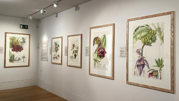
Visual researchers bridged that specific gap. Working alongside botanists at the Royal Botanic Gardens, Kew, illustrators rehydrated pressed fragments, inspected pollen grains under optical microscopes, and painted detailed reconstructions of living plants. Their diagrams showcased floral anatomy in full color: longitudinal dissections of ovaries, magnified views of glandular hairs, and exact transverse cross-sections of seed capsules.
These illustrations functioned as scientific instruments. A researcher working in the Herbarium Library could compare an ambiguous dried specimen collected in South Africa or India with an illustrator’s life-sized rendering of the living plant. By capturing dynamic phenology, from seedling emergence to mature seed-pod explosion, scientific illustrators created reliable diagnostics that pure text descriptions and fragile dried leaves could never achieve alone.

