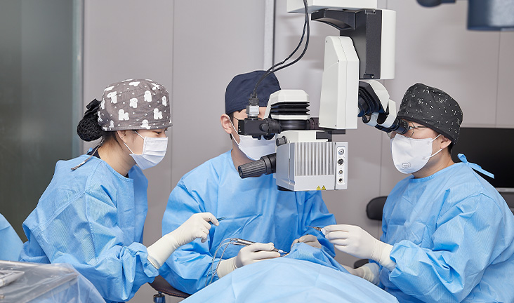
The procedure does not involve cutting into the globe of the eye, lasers, or corneal shaving. Instead, the ophthalmic surgeon works entirely on the external surface beneath the clear conjunctival membrane, manipulating the six delicate extraocular muscles that orchestrate eyeball trajectory.
During the operation, the surgeon accesses the targeted muscle through a microscopic incision. If a muscle pulls too tightly, pulling the eye inward, the surgeon performs a recession, detaching the muscle from its anatomical insertion point and stitching it further back along the sclera to slacken tension. Conversely, if a muscle is lax, a resection or plication shortens the tissue, pulling the eye straight. Precision matters down to fractions of a millimeter: a 1-millimeter variance can alter eye alignment by two to three degrees.
| Surgical Factor | Pediatric Strabismus Protocol | Adult Strabismus Protocol |
|---|---|---|
| Primary Objective | Prevent amblyopia; establish baseline neural binocular pathways. | Eliminate diplopia; recover binocular fusion; expand field of view. |
| Anesthesia Approach | Full general anesthesia exclusively. | General or monitored conscious sedation with local retrobulbar blocks. |
| Suture Technique | Standard absorbable sutures tied permanently during surgery. | Adjustable suture loops fine-tuned post-operatively while awake. |
| Primary Success Rate | 70%, 80% single-operation alignment. | 80%, 88% target alignment using adjustable techniques. |
The introduction of adjustable sutures revolutionized adult outcomes. Instead of permanently securing the muscle while the patient is unconscious, the surgeon secures the repositioned muscle with a slipknot. Hours later, or the following morning, the surgeon assesses the patient’s conscious eye alignment. Using topical numbing drops, the surgeon nudges the suture millimeters forward or backward until the patient confirms that double vision has vanished. This real-time calibration significantly cuts the need for secondary re-operations.

