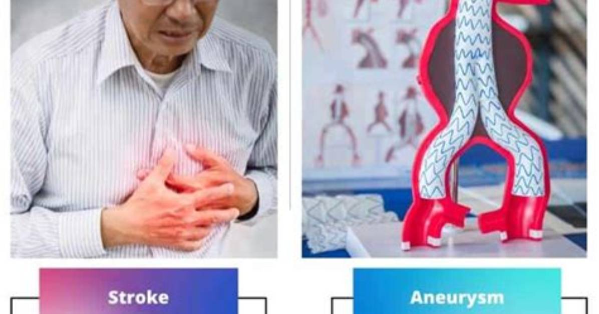
Automated triage algorithms and real-time deep-learning pipelines are dismantling long-standing delays in acute neuroimaging interpretation. In hospitals running integrated imaging platforms, artificial intelligence algorithms analyze non-contrast CT and CTA scans the moment reconstruction finishes, flagging hyperdense subarachnoid blood, quantifying ischemic core volumes, and pinpointing vessel occlusions before the radiologist even opens the primary workstation queue.
These digital screening tools do not replace clinical judgment; they collapse the minutes required to mobilize neurointerventional teams. Whether distinguishing the faint hyperdense signature of a ruptured basilar tip aneurysm from the subtle tissue edema of an evolving middle cerebral artery occlusion, the diagnostic objective remains unchanged. Rapid cross-sectional neuroimaging turns invisible intracranial vascular crises into actionable maps, giving clinicians the exact coordinates needed to preserve viable brain tissue.

