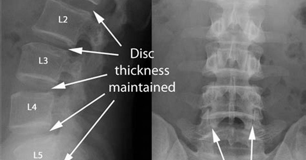
The natural patient reaction to blinding lumbar pain is demanding an immediate scan. Patients assume an MRI or X-ray will pinpoint the precise defect behind their suffering. Clinical practice patterns reveal a different story. The American Academy of Family Physicians consistently advises against routine imaging within the first six weeks of non-specific back symptoms unless red-flag indicators appear.
| Imaging Modality | What It Captures Effectively | Diagnostic Limitations & Risks |
|---|---|---|
| Plain Radiograph (X-Ray) | Vertebral alignment, gross fractures, bony spurs, gross disc height collapse. | Cannot view soft tissue; blind to disc herniation, muscle strains, or nerve inflammation. |
| Magnetic Resonance Imaging (MRI) | Hydration loss, disc extrusion, neural impingement, marrow edema, soft-tissue tearing. | High false-positive rate; highlights age-related changes that may not cause the pain. |
| Computed Tomography (CT) | Complex facet joint anatomy, micro-fractures, structural bone morphology. | Exposes patient to substantial ionizing radiation; inferior soft-tissue contrast compared to MRI. |
Diagnostic imaging frequently uncovers structural anomalies that have zero correlation with the patient's immediate pain. Landmark clinical studies demonstrate that over 30% of pain-free thirty-year-olds display disc bulges on MRI scans, a figure rising to more than 60% in asymptomatic individuals over age fifty. An MRI taken during an acute flare-up often captures what radiologists term "wrinkles on the inside." Labeling these age-appropriate baseline shifts as critical defects can induce kinesiophobia, the debilitating fear of movement that paradoxically delays tissue recovery.

